Remembering Rosalind Franklin, whose photograph was crucial to discovering DNA’s structure
Account subscription benefits alongside Premium Stories, Editorials, Opinions and more. Unlock these with Subscription
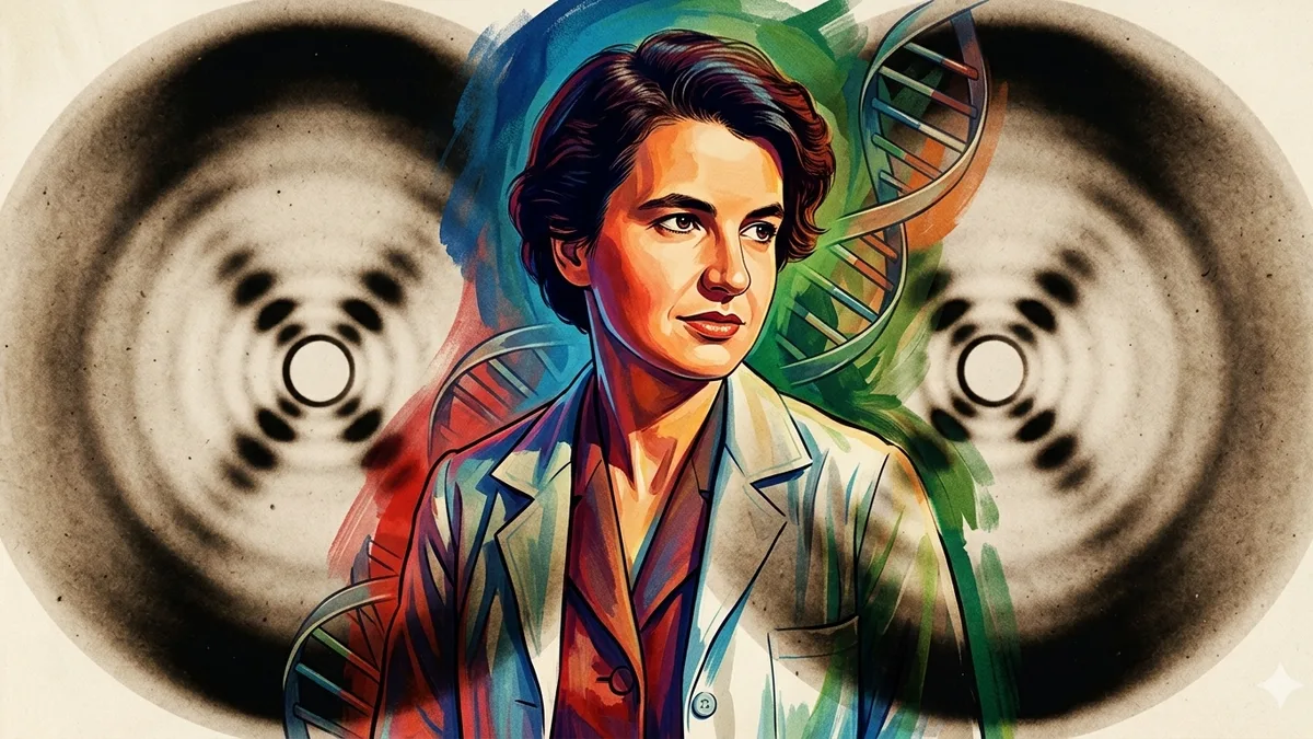
Franklin was a British chemist and X-ray crystallographer. In 1951, she joined the team at King’s College London that were using X-ray diffraction to study the structure of the molecule | Photo Credit: Image generated with AI
For a discipline so wedded to reason and fact as science is, it has fiendishly guarded its gender bias. Over centuries, pioneering women in science have been ignored, their achievements overlooked or usurped by male colleagues, their names left out of scientific publications; they have been underpaid and undervalued, denied promotions and advancements in careers, sometimes consigned to housekeeping roles about the office in order to keep their jobs.
In a compilation of such women in scientific research through the ages, Rosalind Franklin would probably be right on top of the list. It was Franklin’s X-ray diffraction image that lore says, was crucial to discovering the DNA’s double helix structure, enabling advances in genetics and molecular biology, with vast applications in medicine. And yet, when the Nobel Prize for Physiology in 1962 was announced, the names on that list were Watson and Crick, shared with Wilkins. It was decades before her true role in this path-breaking discovery came to be known to the world.
Today, Franklin stands testimony to the power of women, as she is representative of the ‘woman wronged by science’, but not just that – her achievements in science (in other fields) have broken new ground that researchers have since travelled on. As we unveil the achievements of women in science, particularly those whom their present refused to acknowledge, it is important to get her story out there, more so since she was vilified and mocked by the very men who benefitted from her work .
Franklin was a British chemist and X-ray crystallographer. In 1951, she joined the team at King’s College London, biophysicists led by John Randall, and Maurice Wilkins as his deputy (who would later share the Nobel prize), that were using X-ray diffraction to study the structure of the molecule, recount Matthew Cobb and Nathaniel Comfort, two researchers looking into the past of Watson and Crick to craft their biographies, in an article in Nature .
Franklin, had already been trained in this technique, having used it earlier to study the structure of coal, which she advanced by many lengths with her work. Cobb and Comfort detail in their article: “Franklin was able to exploit a discovery that Wilkins had made earlier — DNA in solution could take two forms, what she called the crystalline or A form, and the paracrystalline or B form. Franklin found that she could convert A into B simply by raising the relative humidity in the specimen chamber; lowering it again restored the crystalline A form.” A B form X-ray image it was that provided great clarity on the actual structure of the DNA.
The distorted story of Franklin’s role was spread, thanks in good measure, by Watson’s best selling book The Double Helix .
5News aggregated this summary from the outlet’s public feed. The full article, with all the context, is on www.thehindu.com — the content belongs to The Hindu.